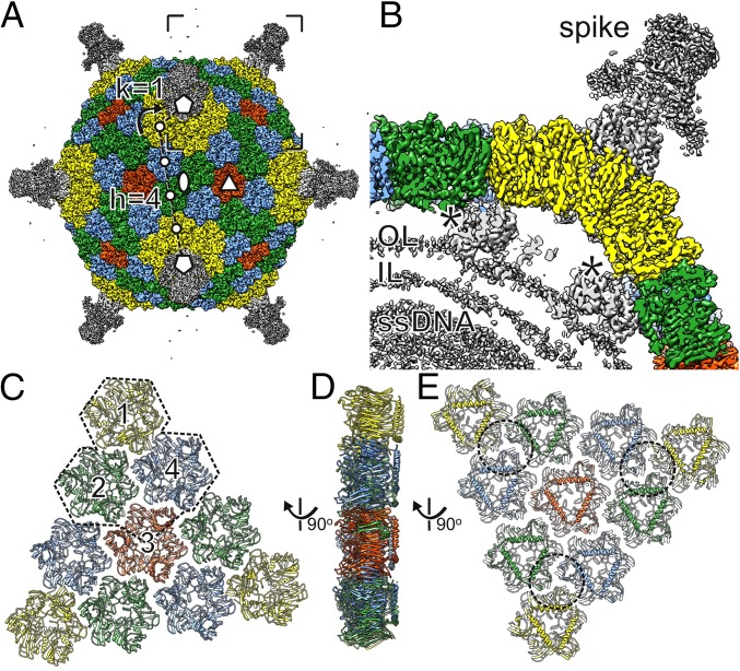
Fig. 2.
3D cryo-EM reconstruction of the FLiP virion. (A) Surface rendering of the FLiP virion. The MCPs are colored yellow, green, blue, and red. Other structural components are colored gray. The icosahedral twofold (ellipse), threefold (triangle), and fivefold (pentagons) axes of symmetry are indicated. The triangulation number T = 21 of the icosahedral lattice of the capsid is calculated as T = h2 + h × k + k2 where lattice indices are h = 4 and k = 1 as indicated. The lattice is right-handed (dextro) as indicated by the curved arrow. (B) A section of virion density is shown from the area indicated in A. Spike structure, minor proteins (asterisks), the outer leaflet (OL) and inner leaflet (IL) of the lipid bilayer, and density corresponding to the ssDNA are indicated. (C) The capsid is composed of 20 faces, each consisting of a group of 10 pseudohexameric MCP trimers (ribbons). Each face is divided in three asymmetric units (one is outlined), each consisting of three MCP trimers (1, 2, 4) and one chain from MCP trimer 3 (in addition to the base domain of the spike). (D and E) Group-of-ten seen from the side (D) and below (E). Approximate footprints of the minor capsid proteins indicated in B are outlined.

