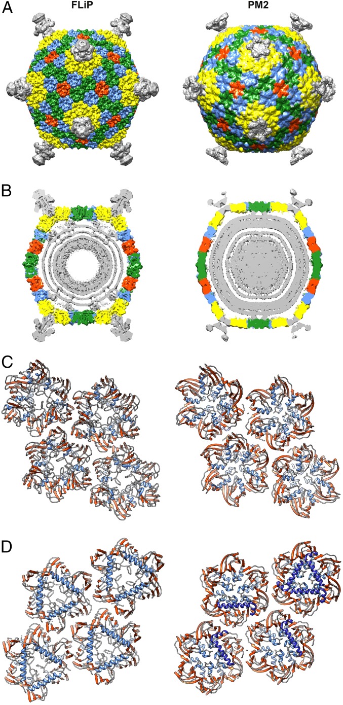
Fig. 4.
Comparison of FLiP and PM2 capsid architecture. (A) Surface-rendered views of FLiP (this study) and PM2 (EMDB accession no. 1082) virion structures. Both maps have been low-pass filtered to 8-Å resolution. MCP trimers are colored yellow, blue, green, and red, depending on their location in the icosahedral capsid. Other densities, including the spikes in the icosahedral vertices, are colored gray. (B) A central section of density is shown for the two virions. Coloring is as in A. Capsid-to-membrane contacts are indicated by arrowheads. In FLiP, these densities have not been assigned. In PM2, they correspond to minor protein P3 (26). (C and D) A ribbon representation of four different types of MCPs seen from outside (C) and inside (D) the virion. In addition to the MCP, α-helices of the PM2 minor protein P3 interacting with the MCP (26) are shown in dark blue.

