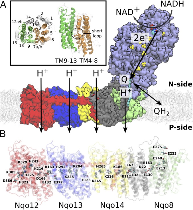
Fig. 1.
Overall structure and function of bacterial complex I from T. thermophilus (PDB ID: 4HEA). (A) Electron transfer from NADH to Q in the hydrophilic domain (in purple) couples to proton transfer across the membrane domain, shown in red (Nqo12), blue (Nqo13), yellow (Nqo14), gray (Nqo11, Nqo10, and Nqo7), and green (Nqo8). (Inset) Transmembrane (TM) helix numbering of the antiporter-like subunits. Symmetry-related transmembrane helices TM4–8 (in orange) and TM9–13 (in green) in Nqo13, and TM15 (in transparent white) from Nqo12. (B) Array of polar and charged residues on the central axis in the membrane domain.

