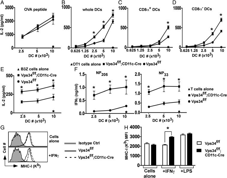
Fig. 3.
Effects of Vps34 deficiency on the classic MHC class I antigen-presentation pathway in DCs. (A) Splenic DCs were pulsed with MHC class I-restricted OVA peptides for 1 h in complete medium and were washed, OT-I T cells were added, and culture supernatants were collected after 24 h to measure IL-2 by CBA. Representative plots from two experiments with four mice per group are shown. (B) Total splenic DCs were loaded intracellularly with OVA protein by osmotic shock, washed, and cultured with 2 × 10⁵ OT-I T cells, and culture supernatants were collected at 24 h for measurement of IL-2. (C and D) Splenic CD8α+ (C) and CD8α− (D) DCs were sorted from the indicated mice, intracellularly loaded with OVA, and cultured with OT-I T cells for 24 h. IL-2 was measured in the culture supernatant. A representative of three experiments is shown. The error bars indicate the means ± SD of triplicate wells. *P < 0.05. (E) BMDCs were loaded with OVA by osmotic shock and cocultured with 2 × 10⁴ B3Z hybridoma cells overnight, and then IL-2 in the supernatant was measured. Representative graphs from two experiments with four mice per group are shown. The error bars indicate the means ± SD of triplicate wells. *P < 0.05. (F) Splenic DCs (2 × 10⁴) were infected with LCMV for 3 h, washed, and cultured with 2 × 10⁵ cells of short-term CD8+ T-cell lines specific for the LCMV-derived nucleoprotein peptides NP205 or NP33 for 36 h. The culture supernatants were collected, and IFN-γ was measured by ELISA. Representative graphs from three experiments with five mice per group are shown. Error bars indicate the means ± SD of triplicate wells. *P < 0.05. (G and H) Total splenic DCs were purified and stimulated with or without 15 ng/mL of IFN-γ or 1 μg/mL of LPS for 16 h. MHC class I (Kb) was measured by flow cytometry. A representative plot (G) and a summary of results pooled from three separate experiments (H) are shown. *P < 0.01.

