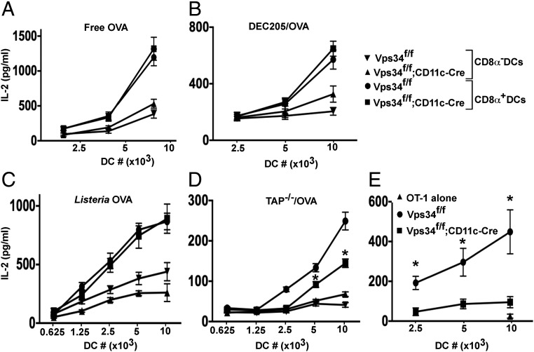
Fig. 5.
Cross-presentation of MHC class I antigens by Vps34-deficient DCs. (A) Splenic CD8α+ and CD8α− DCs (104 cells) were cultured with varying amounts (62.5–250 μg/mL) of free OVA and 2 × 10⁵ OT-I T cells for 24 h. Supernatants were collected, and IL-2 was measured by CBA. (B) Splenic CD8α+ and CD8α− DCs (106 cells) were incubated with biotin-labeled anti-DEC205 antibodies followed by streptavidin-OVA delivery reagent and were cultured in the presence of 2 × 10⁵ OT-I T cells for 24 h. Supernatants were collected for measurement of IL-2 by CBA. (C) Splenic CD8α+ and CD8α− DCs were cultured with OVA-expressing L. monocytogenes and 2 × 10⁵ OT-I T cells for 24 h. Supernatants were used to measure IL-2 by CBA. (D) TAP−/− splenocytes were intracellularly loaded with OVA, sublethally irradiated, and cultured with splenic CD8α+ or CD8α− DCs. OT-I T cells (2 × 10⁵) were added and were cultured for 24 h. Supernatants were collected to measure IL-2 by CBA. In A–D, graphs are representative of three individual experiments with five mice per group. Error bars indicate the means ± SD of triplicate wells. *P < 0.05. (E) Mice were injected with 20 × 106 apoptotic TAP−/− splenocytes loaded intracellularly with OVA by osmotic shock. After 2 h, splenocytes were prepared, and DCs were purified and cultured in the presence of OT-I T cells for 24 h. The culture supernatants were collected to measure IL-2 by CBA. Error bars indicate the mean ± SEM for five mice. *P < 0.05.

