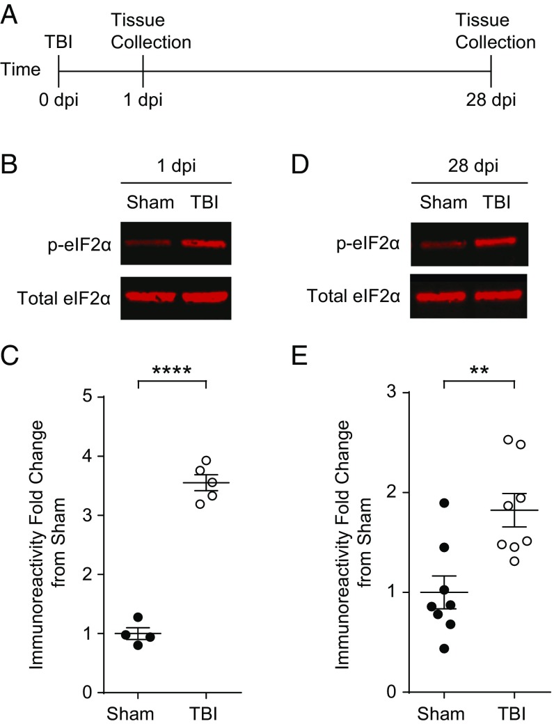
Fig. 1.
TBI-induced increase in eIF2α phosphorylation persists 4 wk after injury. (A) Experimental design scheme. Animals were given a focal TBI by the controlled cortical impact method and the hippocampus ipsilateral to the injury was collected at either 1 dpi or 28 dpi. Sham controls received a craniectomy without a TBI and were analyzed at the same time points. (B) Representative images of p-eIF2α and total eIF2α Western blots from the hippocampi protein samples collected at 1 dpi. (C) Quantification of p-eIF2α to total eIF2α ratio normalized to sham. TBI increases phosphorylation of eIF2α at 24 h postinjury. Data are means ± SEM (n = 4–6 per group, Student’s t test; ****P < 0.0001). (D) Representative images of p-eIF2α and total eIF2α. Western blots from the hippocampi collected at 28 dpi. (E) Quantification of p-eIF2α to total eIF2α ratio normalized to sham. The increase in p-eIF2α in TBI animals persists at 28 dpi. Data are means ± SEM (n = 8 per group, Student’s t test; **P < 0.01).

