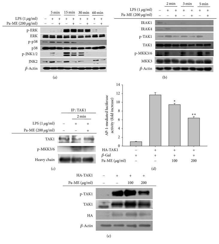
Figure 4.
Effects of Pa-ME on the activation of upstream signaling molecules for AP-1 translocation. (a and b) Phosphorylated or total forms of ERK, p38, JNK1/2, IRAK1, IRAK4, TAK1, MKK3/6, MKK3, and β-actin levels at 5, 15, 30, and 60 min or 2, 3, and 5 min were detected by immunoblotting analysis with phospho-specific or total protein antibodies from total cell lysates. (c) The binding level of p-MKK3/6 to TAK1 at 2 min was detected by immunoprecipitation and immunoblotting analysis of LPS-treated RAW264.7 cells in the presence or absence of Pa-ME. β-Gal, β-galactosidase. ∗P < 0.05 and ∗∗P < 0.01 compared with control group.

