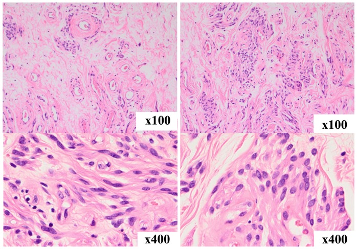
Figure 4.
Histopathological findings. The tumor consisted of sparse collagen fibers and exhibited a higher cellular density around blood vessels. The cells proliferating around blood vessels were oval to spindle-shaped and had acidophilic and fibrous cytoplasm. No mucin deposition was seen in the stroma, and there was no thickening or hyalinization of vascular walls.

