
Figure 2.
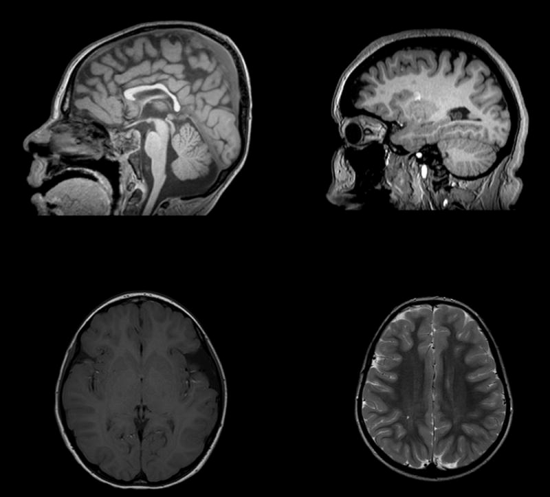
Brain Magnetic Resonance Imaging (MRI) in individuals with loss of function mutations in SHANK3 (A) Sagittal T1 weighted image of subject 9 demonstrating thinning of the corpus callosum (B) Sagittal T1 weighted image of subject 17 with mild cerebral volume loss (C) Axial T1 weighted image of subject 21 with left sylvian fissure arachnoid cyst (D) Axial T2 weighted image of subject 4 with mild T2 hyperintensity of the posterior centrum semiovale.