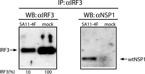
Fig. 1.
NSP1–IRF3 complexes formed in infected cells. IRF3-specific antiserum was used to prepare immunoprecipitates from SA11-4F- or mock-infected Caco-2 cells. The precipitates were analyzed by using Western blot assay with IRF3 or NSP1(C19)-specific antiserum. Intensities (percent) of IRF3 bands were determined with a PhosphorImager and normalized to 100% for IRF3 from mock-infected cells.

