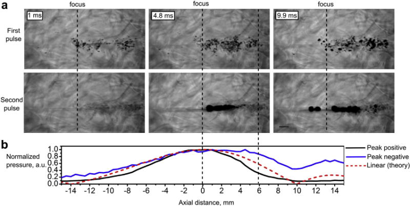
Fig. 5.

A representative set of high-speed camera frames of the PA gel corresponding to the first and second 10-ms pulses, separated by 10 s at 1.5-MHz frequency and high output level. (HIFU is incident from the right, the scale bar is 1 mm.) Boiling does not occur within the first pulse because of prefocal shielding by the cavitation bubbles. However, the first pulse depletes most cavitation nuclei and boiling starts within the second pulse at 4.8 ms. The normalized axial distribution of peak positive (black line) and peak negative (blue line) pressures recorded by FOPH in water in the shocked regime and the theoretically predicted axial pressure distribution in the linear regime (bottom plot, dashed red line). The distributions are shown in the same scale as the high-speed camera images (0 corresponds to the focus of the transducer). PA = polyacrylamide; HIFU = high-frequency focused ultrasound; FOPH = fiber optic probe hydrophone.
