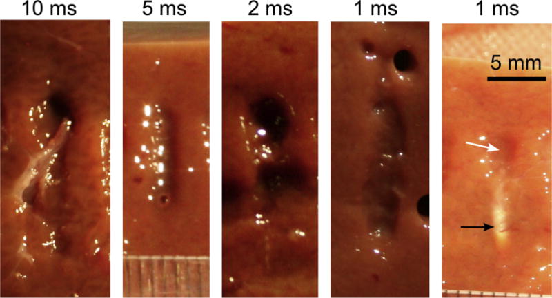
Fig. 7.

BH lesions produced in bovine liver at the frequency of 1.5 MHz and different pulse durations, while keeping the number of pulses delivered per focal spot and duty factor the same (30 and 1%, correspondingly). The smaller pulse duration implies higher shock amplitude at the focus, so that boiling occurs at each pulse. Peak negative in situ focal pressure is also increased, and at 1-ms pulse duration it is estimated as 14.5 MPa. At 1-ms pulse duration, ghost lesions are occasionally formed (rightmost panel) indicating the onset of prefocal cavitation that shields the focus. As also shown in Fig. 6b, the ghost lesion consists of a narrow thermally denatured area around the focus (black arrow) and an area of mechanically disrupted tissue prefocally (white arrow). (HIFU is incident from the top of the images.) BH = boiling histotripsy; HIFU = high-intensity focused ultrasound.
