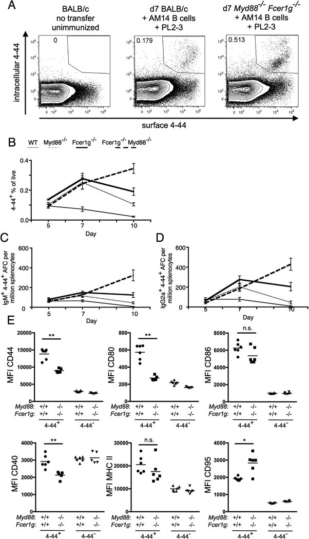
Figure 1.

FcRγ and MyD88 control contraction of the EF RF response. BALB/c or Myd88-deficient BALB/c or Fcer1g-deficient BALB/c or MyD88-deficient Fcer1g-deficient BALB/c mice were sacrificed on day 5, 7, or 10 following transfer of purified AM14 sd-Tg B cells and administration of PL2-3 as indicated in Materials and Methods. (A) Representative staining of surface and intracellular 4-44 double positive cells in indicated recipients at d7 following transfer and immunization. (B) Surface and intracellular 4-44 double positive cells as gated in (A) were quantitated by flow cytometry. (C-D) Splenic 4-44+ AFC of IgM (C) or IgG2a (D) isotype were measured by ELISpot. At least 4 mice per group and 2 independent experiments per time point were compiled. Data are represented as means +/- SEM. (E) Expression of indicated activation markers on 4-44+ or 4-44- B cells at d7 in the indicated recipients was determined by flow cytometry. Statistical comparisons by Mann-Whitney two-tailed test, * p<0.05; ** p<0.01
