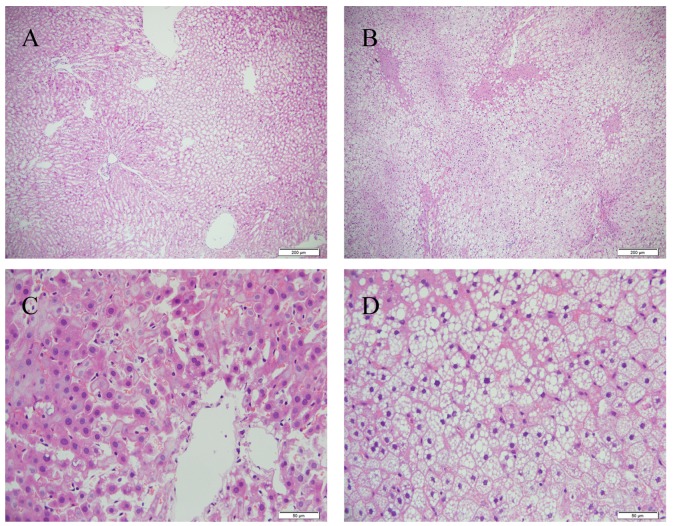
Figure 1.
Hepatic pathology. Liver sections from the Con and HFD groups were stained with hematoxylin and eosin. Representative images from (A) Con and (B) HFD groups at ×100 magnification. Representative images from (C) Con and (D) HFD groups at ×400 magnification. Fat droplets, ballooned hepatocytes and inflammatory cells are present in the HFD group. HFD, high fat and high cholesterol diet; Con, control; HFD, high fat and high cholesterol diet.

