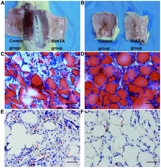
Figure 1.
Grafted adipose tissue was removed at 5 weeks. The left sides were the control group, and the right sides were the BoNTA group (A and B). After Oil Red O staining, cellular integrity was assessed as grade 2.50±0.548 in the control group (C), and cellular integrity was assessed as grade 4.67±0.516 in the BoNTA group (D). The red arrows indicate apoptotic cells; visibly less apoptosis was detected in the BoNTA group (F) than that in the control group (E) by TUNEL staining. Cellular integrity parameter was graded on a semi-quantitative scale of 0 to 5 as follows: the percentage of normal shape adipocytes was 1 (< 5%), 2 (5–25%), 3 (25–50%), 4 (50–75%), 5 (>75%). (C–F) scale bar, 50 µm. BoNTA, botulinum toxin A.

