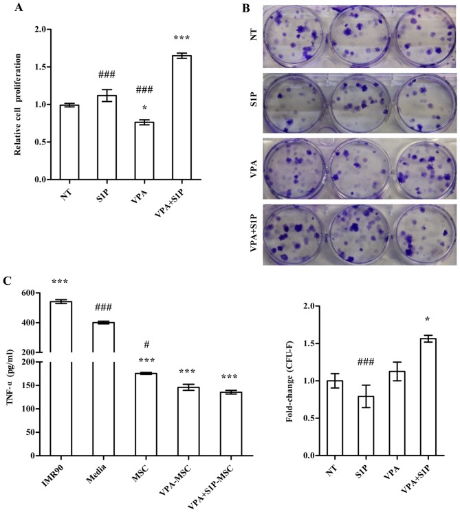
Figure 4.
Enhanced therapeutic potency of umbilical cord-derived mesenchymal stem cells (UC-MSCs) primed with valproic acid (VPA)+sphingosine-1-phosphate (S1P). (A and B) Cell proliferation analysis [(A) n=4)] and colony-forming unit-fibroblast (CFU-F) assay [(B) n=6)] of UC-MSCs primed with VPA (0.5 mM) or S1P (50 nM) alone as well as VPA+S1P for 24 h. For the CFU-F assay, 600 cells were seeded into 6-well culture plates and cultured for 14 days, and the number of colonies was quantified. Representative stained colonies of adherent cells are shown in the right panel. (C) Quantification of TNF-α protein (n=4) secreted from a murine alveolar macrophage cell line stimulated with LPS for 5 h in the absence or presence of conditioned medium (CM) harvested from the indicated cells. All data are presented as means ± SEM, *p<0.05, **p<0.01 and ***p<0.001 compared with non-treated (NT) umbilical cord blood (UCB)-MSCs; #p<0.05 and ###p<0.001 compared with VPA+S1P primed cells, one-way ANOVA test ANOVA with Bonferroni post-test.

