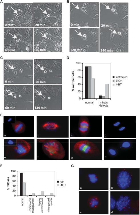
Figure 1.

Wee1 mutant cells display mitotic aberrations. (A–C) Time-lapse images of dividing Wee1Co/−;TM-Cre MEFs either treated with ethanol (ctr) (A) or 4-hydroxytamoxifen (4-HT) (B, C). Representative images show either normal mitosis (A) or mitotic cells that cannot complete mitosis (B) or cells that withdraw from mitosis (C). (D) Summary of mitotic abnormalities in Wee1 mutant MEFs during live imaging. (E) Tamoxifen inducible Wee1 knockout MEFs either in the absence (a, b, c, d) or presence (e, f, g, h) of 4-HT were stained with antibodies against α-tubulin (red), Aurora-B (green) and 4′,6-diamidino-2-phenylindole (DAPI; blue). (F) Quantification of the mitotic defects observed in Wee1-deficient cells after treatment with 4-HT. (G) Wee1 knockout MEFs either in the absence (a, b) or presence (c, d) of 4-HT were stained with pericentrin (red) and DAPI (blue).
