Figure 5.

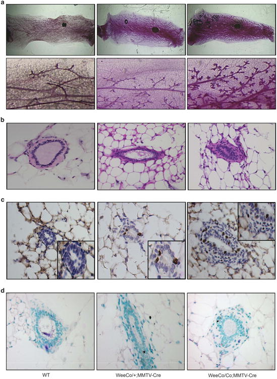
Conditional deletion of Wee1 in mammary gland caused increased cellularity. (a) Whole-mount imaging of mammary glands from 6-month-old virgin WT (left), Wee1Co/+;MMTV-Cre (middle) and Wee1Co/Co;MMTV-Cre mice (right). Boxed areas are enlarged and placed underneath. (b–d) Hematoxylin and eosin stained sections (b), BrdU-positive cells (c) and TUNEL-positive nuclei (d) in 6-month-old virgin WT (left), Wee1Co/+;MMTV-Cre (middle) and Wee1Co/Co;MMTV-Cre mice (right). Boxed areas in c are enlarged areas showing BrdU-positive cells.
