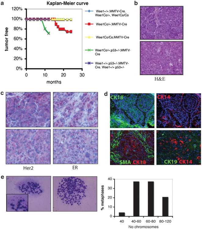
Figure 7.

Tumor formation in mice carrying mammary-specific deletion of Wee1. (a) Kaplan–Meier survival curve of mice with different genotypes as indicated. (b) Representative hematoxylin and eosin stained (H&E) slides from mammary tumors from two Wee1Co/+;MMTV-Cre mice. (c) Sections from mammary tumors from Wee1Co/+;MMTV-Cre mice were stained with antibodies against HER2 and ER by immunohistochemistry. (d) Indirect immunofluorescence in mammary tumors from Wee1Co/+;MMTV-Cre mice with antibodies against SMA, CK14, CK18 and CK19. (e) Metaphases of cell lines developed from primary tumors showing double minute chromosomes (left) and polyploidy (middle). Quantification of chromosome numbers in Wee1-deficient tumor cells is shown (right).
