
Figure 2.
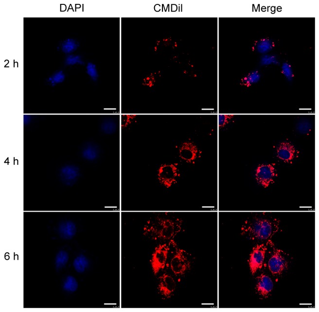
Internalization of BMSC-EVs by HUVECs. HUVECs were incubated with CM-Dil (red)-labeled BMSC-EVs for 2, 4 and 6 h. Cell nuclei were stained with DAPI (blue). Scale bar, 20 µm. BMSC-EVs, bone marrow mesenchymal stem cell-derived extracellular vesicles; HUVECs, human umbilical vein endothelial cells.