Figure 3.
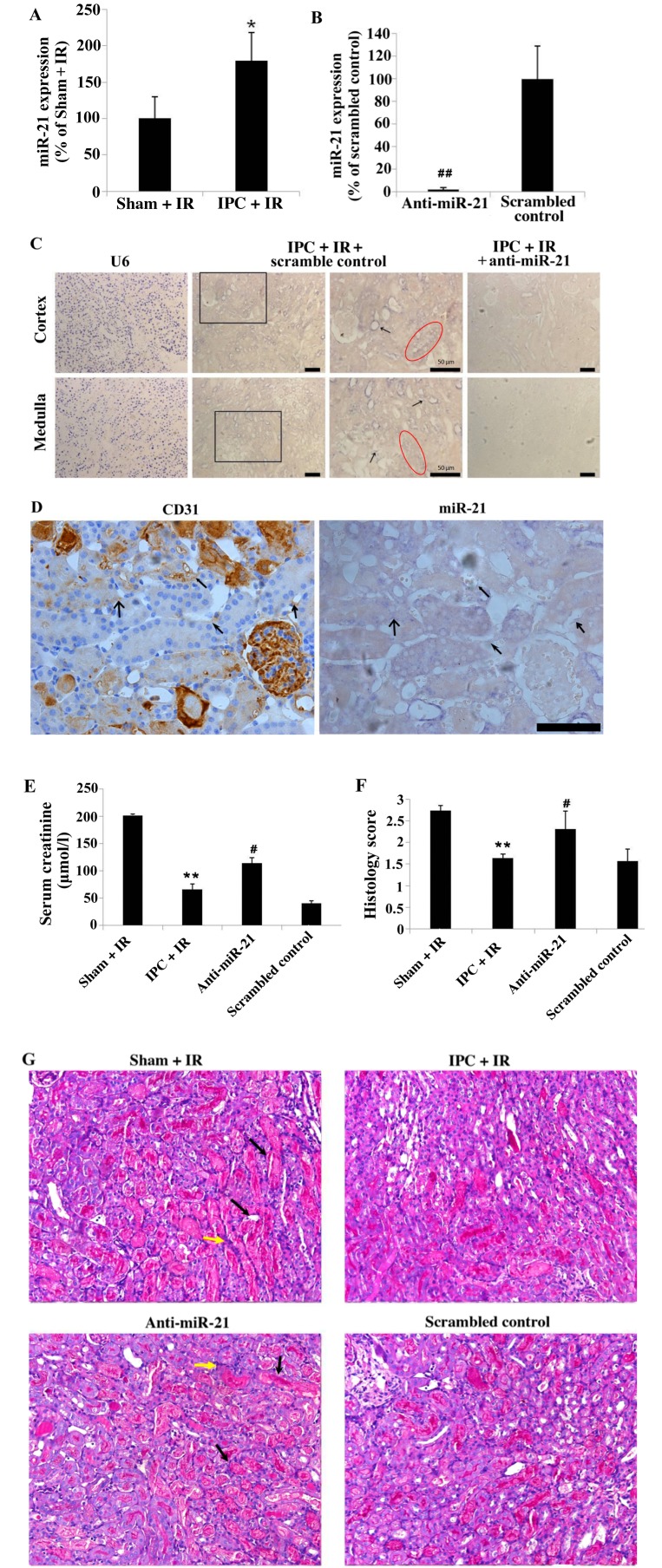
Delayed IPC increased miR-21 expression in renal tubular and in endothelial cells, which was inhibited by locked nucleic acid-modified anti-miR-21 oligonucleotide treatment. (A) miR-21 expression 24 h following IR was increased in kidneys exposed to IPC compared with Sham + IR mice. (B) Expression of miR-21 in the renal tissue 24 h following IR was inhibited by treatment with anti-miR-21 administered at the time of IPC, compared with mice treated with the scrambled control oligonucleotides. (C) Representative images of U6 (positive control) and miR-21 expression in mouse kidney sections by in situ hybridization. The increase of miR-21 expression was notable in vascular endothelial cells (arrows) of IPC + IR + scrambled control-treated mice in addition to the renal tubular epithelial cells (red circle). Magnification, ×20; scale bar, 50 µm. (D) miR-21 expression in vascular endothelial cells, which were marked by CD31 staining and is indicated by the similar shaped arrows in the two images. (E) Serum creatinine and (F) histology score 24 h following IR were attenuated by delayed IPC, wherease miR-21 knockdown in mice exposed to delayed IPC significantly worsened renal IR injury. (G) Histopathological changes of mice kidney sections from each group. Representative periodic acid-Schiff-stained micrographs in the corticomedullary junction. Magnification, ×20. Black arrow indicates cast formation and yellow arrow indicates infiltration of inflammatory cells. Data are presented as the mean ± standard error of the mean. n=6 per group; *P<0.05 and **P<0.01 vs. Sham + IR group; #P<0.05 and ##P<0.01 vs. scrambled control group. IPC, ischemic preconditioning; IR, ischemia/reperfusion; miR-21, microRNA-21; U6, U6 small nuclear RNA.

