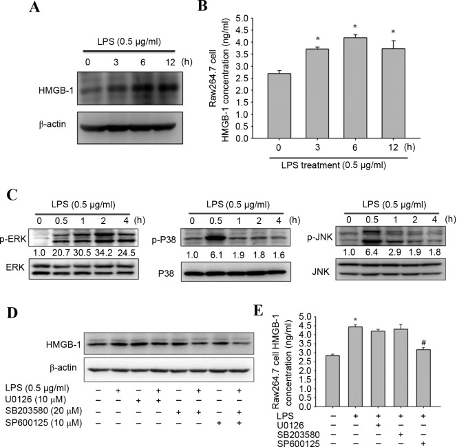
Figure 3.
Effects of LPS on HMGB-1 expression and mitogen activated protein kinase signaling pathway proteins. RAW264.7 macrophages were treated with 0.5 µg/ml LPS for 0, 3, 6 or 12 h. (A) Representative western blot images of HMGB-1 protein expression levels. ß-actin served as an internal control. (B) Quantification of ELISA assay to determine HMGB-1 secretion. (C) RAW264.7 macrophages were treated with 0.5 µg/ml LPS for 0, 0.5, 1, 2 or 4 h. Representative western blot images of ERK 1/2, JNK, p38, p-ERK, p-JNK and p-p38 protein expression levels. Total Erk, total p38 and total JNK were the loading controls. (D) Representative western blot images and (E) ELISA assay results of HGMB-1 levels in macrophages following pretreatment with 10 µM U0126, 20 µM SB203580 or 10 µM SP600125 for 30 min, and incubation with 0.5 µg/ml LPS for 6 h. Data are expressed as the mean ± standard deviation (n=3). *P<0.05 vs. untreated group; #P<0.05 vs. LPS-treated group. LPS, lipopolysaccharide; p, phosphorylated; ERK, extracellular signal regulated kinase; JNK, c-Jun N-terminal kinase; HMGB-1, high mobility group box-1.

