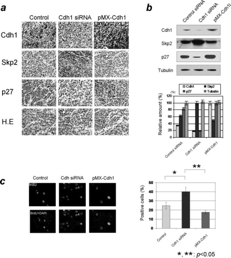
Figure 5. Characterization of the tumors from xenograft mouse study.
a) Immunohistochemical analysis of Cdh1/APC-Skp2-p27 axis in sectioned tumors from the xenograft study. Tumors from Cdh1-overexpressed breast cancer cells were broadly stained positive for Cdh1 and p27 while the cells were mostly negative for Skp2. On the contrary, tumors from Cdh1-knockdown cells appeared to be negative for Cdh1 and p27 while significantly positive for Skp2. b) Evaluation of immunoblotting results using the resected tumors from the xenograft mouse study. Levels of Skp2 markedly increased while levels of Cdh1 and p27 decreased from tumors derived from Cdh1-depleted cells. Levels of Skp2 decreased while levels of p27 increased in tumors from Cdh1-knockdown cells. c) Measurement of S-phase population of the tumor grown in the nude mouse. The population of BrdU positive cells was elevated in the tumors derived from Cdh1-knockdown breast cancer cells.

