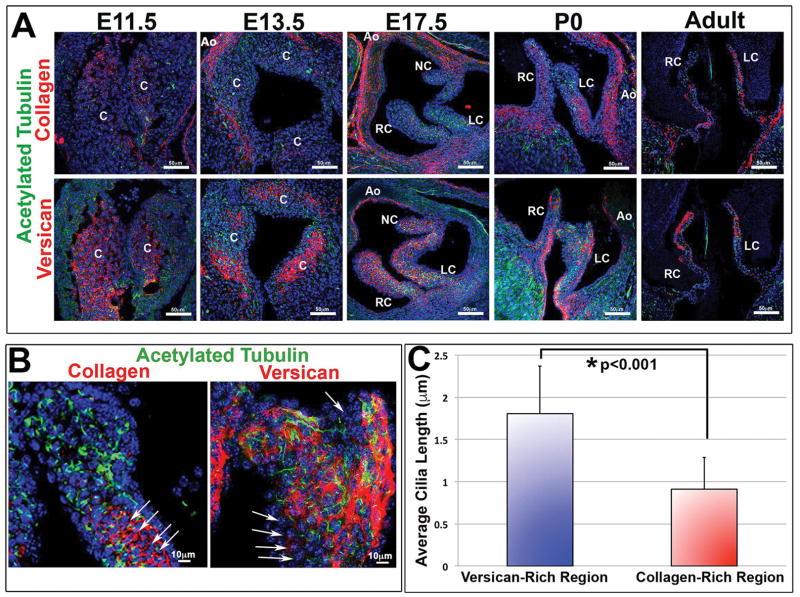
Figure 2. Correlation of primary cilia with versican expressing microenvironments.
(A) IHC for the ciliary axoneme (green), collagen (red, top) versican (red, bottom), and nuclei (blue) show spatial/temporal expression of cilia throughout development. Cilia are predominantly expressed in proteoglycan-rich zones and scant in regions of high collagen expression. C= conal cushions, RC= right-coronary, LC= left-coronary, NC= non-coronary, Ao= aorta. (B) High magnification images of axonemes (green) and collagen/versican (red). Arrows depict collagen rich regions expressing shortened cilia. (C) Quantification of cilia length in both versican and collagen rich regions shows decreased average cilia length in collagen rich regions when compared with versican rich regions, p<0.001 Students t-test.

