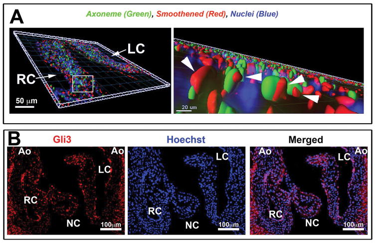
Figure 3. Active Hedgehog signaling in aortic valve interstitial cells in vivo.
(A) Three-dimensional reconstruction of IHC stain at postnatal day 0, shows smoothened (red), acetylated tubulin—cilia axoneme (green), and Hoechst--nuclei (blue). High magnification (right) shows smoothened (arrowhead pointing to smoothened staining) on the axoneme of the cilia indicative of active hedgehog signaling. (B) IHC of P0 aortic cusps showing Gli3 expression.

