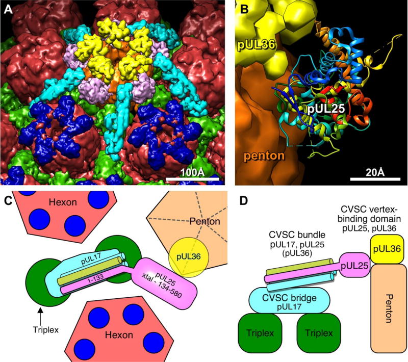
Figure 5. Organization of the CVSC molecule and its binding partners.
(A) Surface view above the penton (brown) including hexons (red), triplex (green), VP26 (dark blue) CVSC trimer consisting of pUL17, the N-terminal domain of pUL25 and the C-terminus of pUL36 (light blue), as well as the C-terminal domain of pUL25 (pink), and the pUL36 density above the penton (yellow). (B) Atomic model of the HSV-1 pUL25 C-terminal fragment (residues 134–580; PDB 2F5U8), fit into the virion capsid cryoEM density map. Note regions of pUL25 that may be contact points with pUL36. Model of the CVSC subunit organization viewed from above (C) and from the side (D) of the penton.

