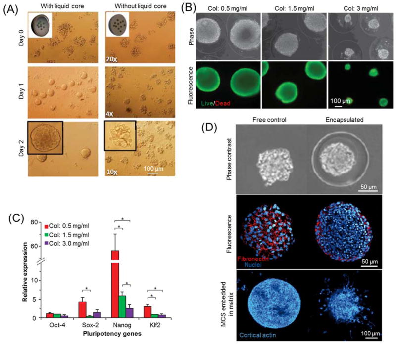
Fig. 6.
Hydrogel microencapsulation for cell growth and proliferation. (A) Typical bright field images of mouse embryonic carcinoma cells encapsulated in alginate hydrogel microcapsules with or without liquid core. Image is reprinted and recreated with permission from reference 78. (B) Phase contrast and fluorescence images of mouse embryonic stem cells (ESCs) encapsulated in collagen core-alginate shell microcapsules with various collagen concentrations. (C) Quantitative RT-PCR of ESC spheroids formed in various core ECM. Images (B–C) are reprinted and recreated with permission from reference 80. (D) Phase contrast and Immunofluorescence staining micrographs of multicellular spheroids (MCS) in liquid core-alginate shell microcapsules and unencapsulated spheroids as free controls. Image is reprinted and recreated with permission from reference 173.

