
Figure 3.
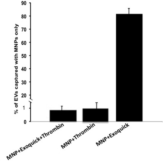
Comparison of different protocols for EV isolation. CD9+/CD41+ EVs were isolated from PPP by capture with MNPs coupled to anti-MHC-I antibodies and the numbers of captured vesicles were evaluated. For each sample, isolation was performed under four conditions: EVs were captured with MNPs (i) from untreated PPP, (ii) from PPP treated with thrombin, (iii) from PPP treated with thrombin and ExoQuick, and (iv) from PPP treated with ExoQuick. To exclude the effects of the donor-to-donor variability, for plasma from each donor, the number of captured EVs was normalized by the number of EVs captured from untreated PPP. Presented are means ± SEM. EV: extracellular vesicle; CD: cluster of differentiation; PPP: platelet-poor plasma; MNP: magnetic iron oxide nanoparticle; MHC-I: major histocompatibility complex class-I; SEM: standard error of the mean.