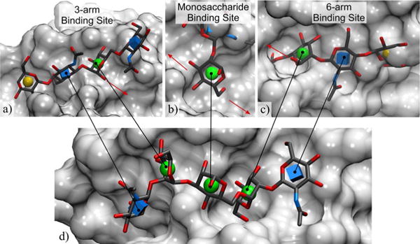
Figure 3.

Structure-based rationalization of the observed binding specificity of ConA towards the arrayed glycans; a) only the weaker 3-arm binding site tolerates linear core m1 trisaccharides and branched glycans, which do not display affinity for ConA on the array; b) the core Man does not tolerate substitution at the 2-position of the bound Man; c) the 6-arm binding site does not tolerate substitution at the 4-position of the bound GlcNAc, nor at the 6-position of the Man, which is consistent with the binding specificity of ConA amongst the arrayed glycans; d) the co-complex of GlcNAcβ1-2Manα1-3[GlcNAcβ1-2Manα1-6]Manα with ConA from PDB ID 1TEI[34] shows the relative position of the 3-arm,6-arm, and monosaccharide binding sites. Glycans represented in 3D-SNFG icon mode,[37] the protein surface is in light grey, red arrows indicate the position of the neighboring binding site(s).
