
Figure 2.
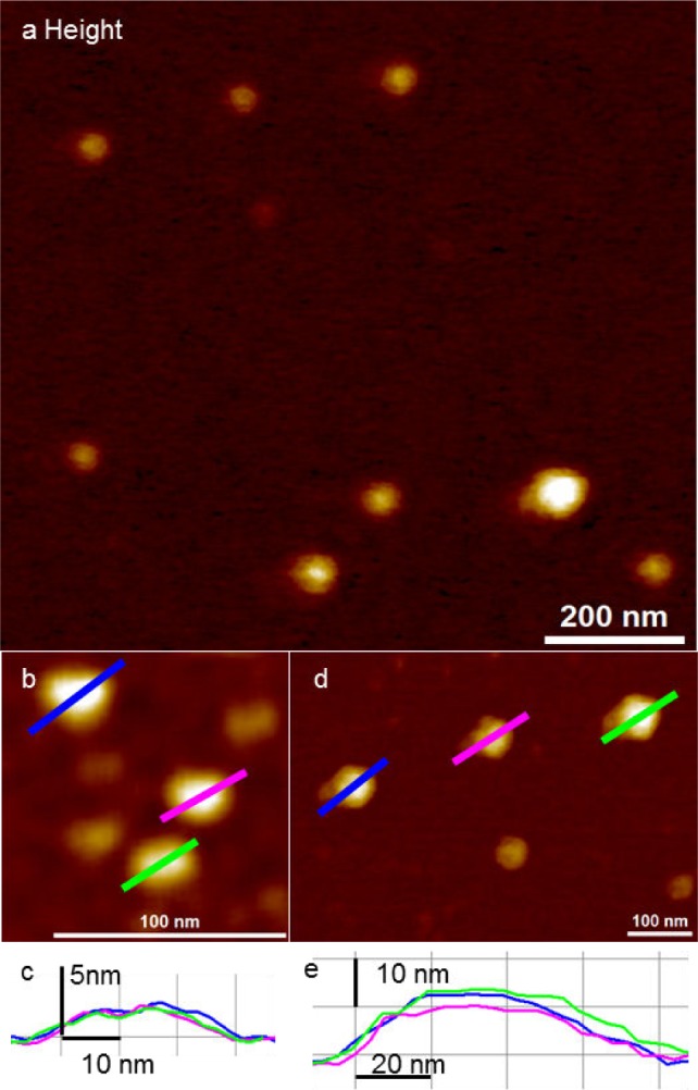
Detection of IA exosomes by peak force AFM imaging in fluid; (a) representative topographic image of exosomes showing varying vesicle size; (b) close-up of smaller IA exosomes. The smaller IA exosomes were 22.5 ± 3.5 nm in diameter and 1.8 ± 0.3 nm in height; (c) cross-section of (b) shows IA exosome dimensions to be approximately 30 nm in diameter and with a height of 2.3 nm; (d) close-up of larger IA exosomes. The dimensions of bigger IA exosomes are a diameter of 64.6 ± 9.2 nm and a height of 8.1 ± 3.1 nm; (e) the cross-section of (d) show IA exosome dimensions of approximately 80 nm in diameter and a height of 12 nm.