
Figure 5.
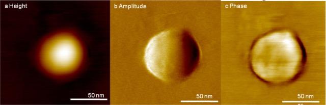
High-resolution images of UC exosomes obtained by AFM imaging in air; (a) representative topography, (b) amplitude and (c) phase images of exosomes. Inset (a) is the cross-section of the exosome for the height channel. The dimensions of the exosome in (a) are a diameter of 79 nm and a height of 6.2 nm. The phase channel shows the smooth surface of the exosome.