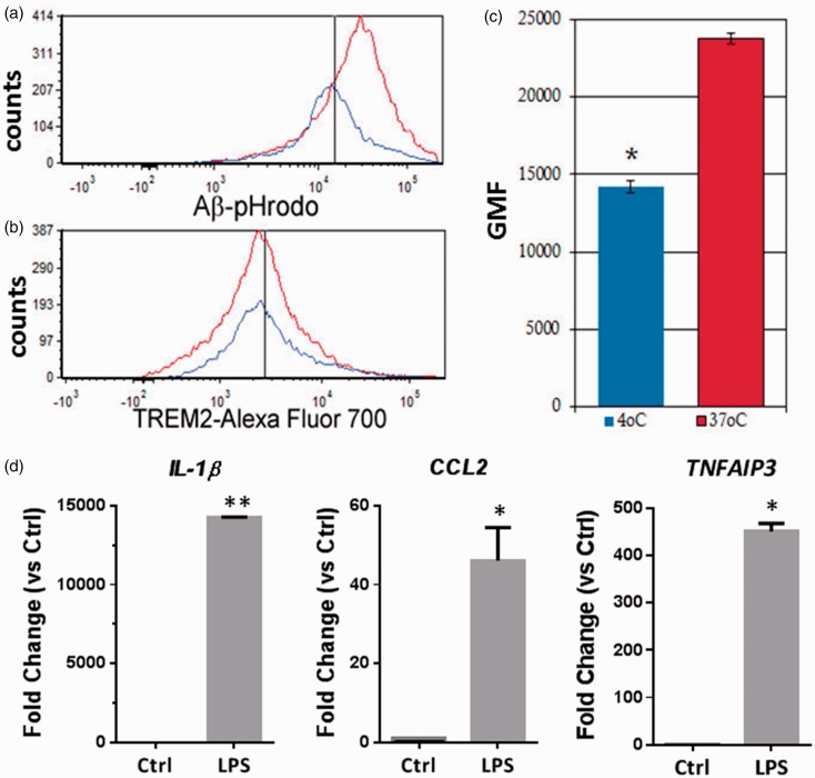
Figure 4.
ScMglia expressing TREM2 internalize Aβ and respond to LPS. (a) Phagocytosis of Aβ was determined by a shift in cellular signal at 37℃ versus signal at 4℃. After gating data based on TREM2 expression, H1 ES-derived microglia-like cells show a positive shift in Aβ fluorophore signal at 37℃ compared with the same cells at 4℃, suggesting the presence of phagocytic internalization of Aβ in TREM2+ differentiated cells. (b) No significant difference was observed with respect to TREM2 expression between Aβ-pHrodo-treated microglial-like cells at 4℃ and those incubated with labeled Aβ at 37℃. Black vertical lines in (a) and (b) represent approximations of the average of the geometric means of fluorescence for cells incubated at 4℃. (c) Quantification of shift in fluorescent signal, indicating phagocytosis of Aβ-pHrodo proteins by TREM2+ H1 ES-derived microglia-like cells. Bars indicate average of geometric means of fluorescence from three independent experiments. (d) Expression of NFkB responsive genes, IL-1β, CCL2, and TNFAIP3 is induced in TREM2+ ES cell-derived ScMglia cells after treatment with LPS. ScMglia were first sorted for TREM2 expression, cultured for 24 hr followed by 24 hr incubation with 50 ng/ml LPS (Student’s t-test, ** p < .01; * p < .05).

