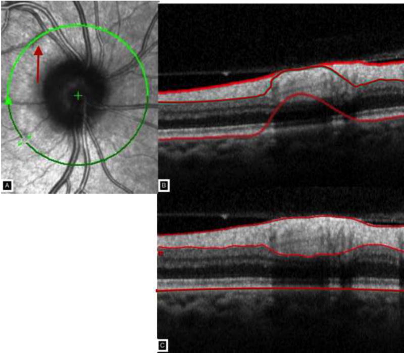
Figure 2.

Panel A shows the circle scan intercepting a retinal vessel for approximately one clock hour creating a shadow and segmentation artifact shown in Panel B. Panel C shows the manual delineation of the nerve fiber layer thickness.

Panel A shows the circle scan intercepting a retinal vessel for approximately one clock hour creating a shadow and segmentation artifact shown in Panel B. Panel C shows the manual delineation of the nerve fiber layer thickness.