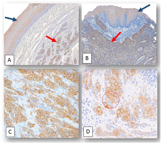
Figure 1. ORF1p expression in normal esophagus and squamous cell carcinoma.

A) and B) Representative photomicrographs depicting LINE-1 ORF1p expression in normal esophageal tissue (black arrows) and in esophageal squamous cell carcinoma cases (red arrows): A) and B) Normal esophageal tissue adjacent to squamous cell carcinoma from two distinct individuals stained with LINE1 ORF1p (final magnification x100). C) and D) are photomicrographs showing a squamous cell carcinoma case where peri-nuclear staining patterns manifest for ORF1p. C) Final magnification x100. D) The same case as C) with a zoomed-in image of ORF1p peri-nuclear staining accentuation (within red circle). Final magnification x160.
