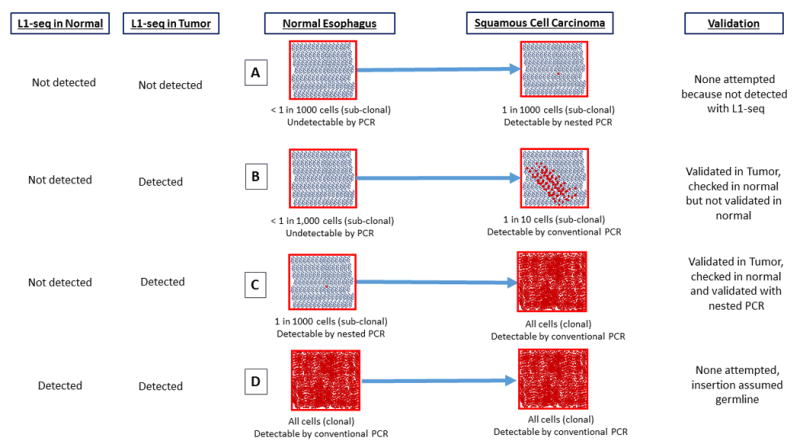
Figure 4. Acquisition, Detection, and Validation of sub-clonal and clonal somatic insertions in L1-seq.

This diagram details the sensitivity of L1-seq with regard to detecting somatic insertions at differing levels of clonality in a tissue and different scenarios by which a somatic insertion could become amplified in a tumor. (A) An insertion present at a very low frequency (less than one in a thousand cells) in the normal esophagus and at an undetectable level in the tumor when evaluated by L1-seq. (B) An insertion at an undetectable level in the normal esophagus and a sub-clonal but detectable level in the tumor. (C) An insertion at undetectable level for L1-seq in the normal esophagus which is clonal in the tumor. (D) An insertion which is clonal in both the normal esophagus and the tumor. This insertion is not tested by PCR because it is presumed to be germline.
