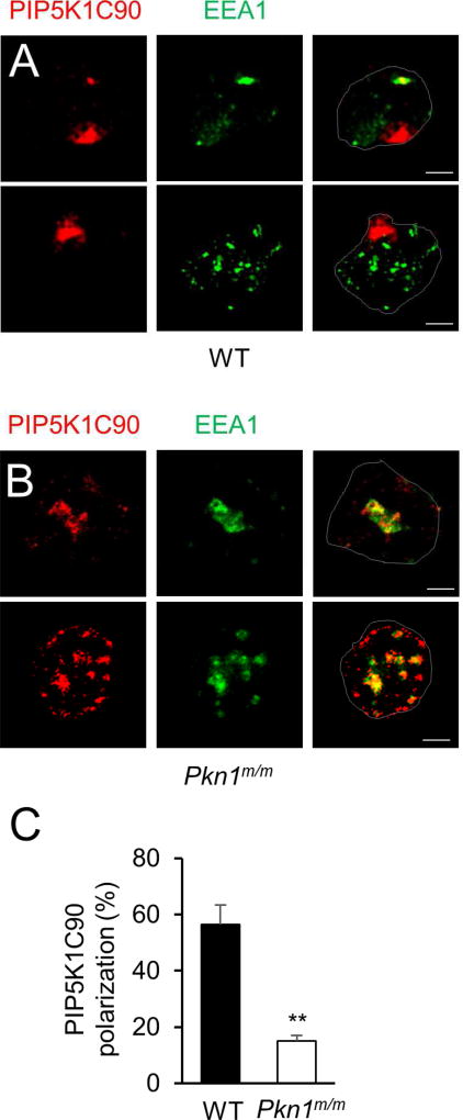
Figure 2. PKN1-deficiency impairs PIP5K1C90 polarization in neutrophils.
A–B. Mouse neutrophils transfected with HA-PIP5K1C90 were placed on Fb-coated coverslips for 30 min. They were then fixed and stained by anti-HA and anti-EEA1 antibodies followed by secondary antibodies conjugated with Alexa633 (colored in red) and Alexa488 (colored in green). Cell contours are outlined and scale bars are 2.5 µm for all of the figures.
C. Quantification of the effect of PKN1-deficiency on PIP5K1C90 polarization. The quantification was done from three independent observations, and more than 20 cells were examined in each observation. Data are presented as means ± SD (**, p<0.01, Student’s t-Test).

