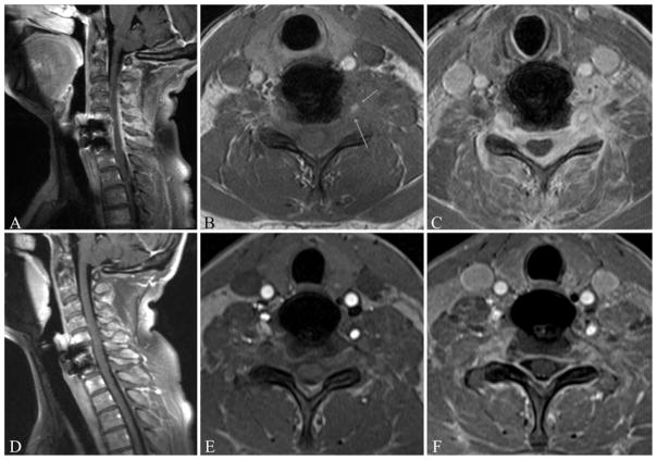
Fig. 4.
Sagittal postcontrast (A) and axial precontrast (B) and postcontrast (C) T1-weighted MR images obtained at the initial presentation (prior to initiation of treatment). Sagittal postcontrast (D) and axial precontrast (E) and postcontrast (F) T1-weighted MR images obtained 6 months after initiation of steroid therapy. The enhancing paraspinal and epidural mass seen in the initial images (A–C) shows stable improvement after 6 months of treatment (D–F). Arrows indicate involvement of the left vertebral artery.

