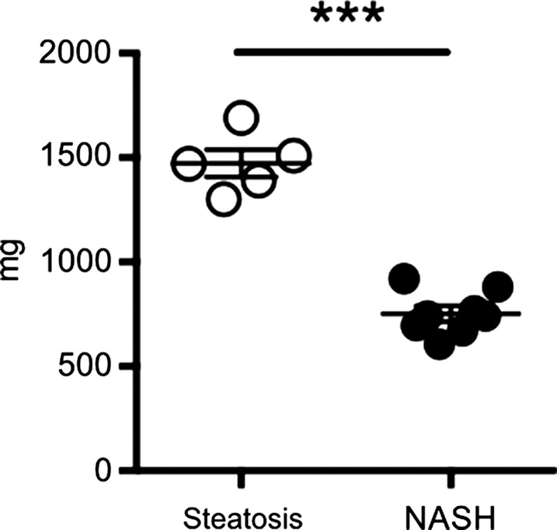
Figure 3d:

Db/db mice fed MCD diet developed NASH per histologic criteria. (a) Hematoxylin-eosin–stained liver sections show markedly increased steatosis, lobular inflammation, and hepatocyte ballooning in NASH compared with steatosis (left: 50×, bar = 250 μm; and right: 200× magnification, bar = 100 μm). (b) Histologic quantification of steatosis in NASH compared with steatosis. (c) Body weight decreased continuously on MCD diet, whereas control mice gained weight over the 4-week study period. (d) Liver weight decreased in these mice. (e) NAFLD activity score was higher in mice fed MCD diet compared with control mice. ** P < .01, *** P < .001. All data are mean ± standard error of the mean.
