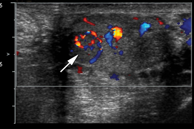
Figure 12b.
Splenogonadal fusion in a 29-year-old man with a painless, palpable left testicular mass, found during workup for infertility. (a, b) Gray-scale US (a) and color Doppler (b) images of the left testis show a hypoechoic mass (arrow in a) with internal flow (arrow in b). (c) Photograph of the gross pathology specimen shows a mass (arrow) representing splenic tissue.

