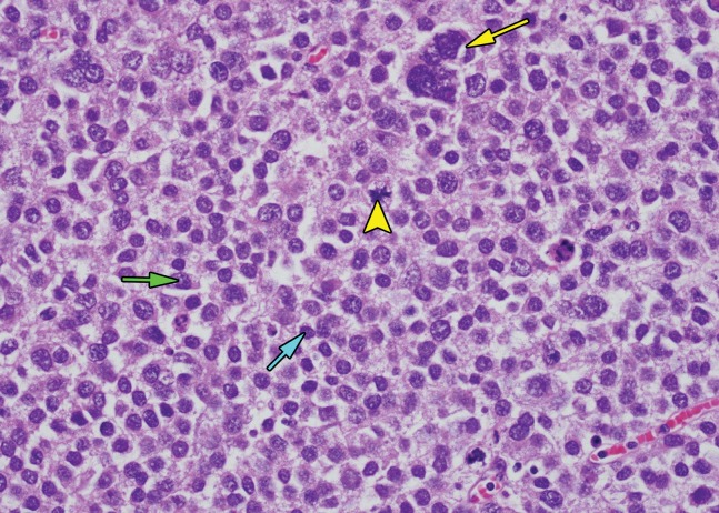
Figure 16c.
Spermatocytic tumor in an 85-year-old man with an enlarging, painless right testicular mass. (a) Panoramic gray-scale US image shows a markedly enlarged heterogeneous right testis (arrow) adjacent to the normal left testis (*). (b) Photograph of the gross pathology specimen shows the heterogeneous appearance of the tumor (arrow). (c) High-power photomicrograph shows the characteristic small cells (blue arrow), intermediate cells (green arrow), and giant cells (yellow arrow) of spermatocytic tumor. The intermediate cells show a spireme-like chromatin distribution. Mitoses (arrowhead) are also present. (H-E stain.)

