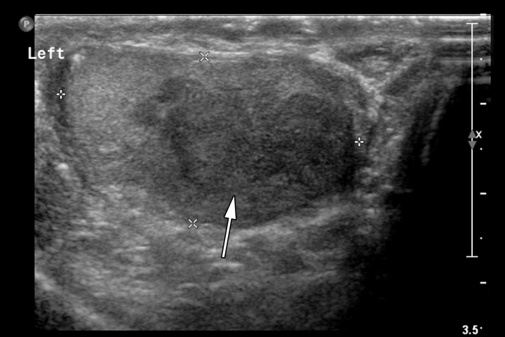
Figure 6e.
Retroperitoneal adenopathy in a 45-year-old man with left back pain. (a) Coronal contrast-enhanced CT image shows left para-aortic adenopathy (white arrow) just below the level of the left renal artery (black arrow). (b–d) Coronal T2-weighted single-shot fast spin-echo (SSFSE) (b), axial T2-weighted SSFSE (c), and axial diffusion-weighted (d) MR images show the retroperitoneal adenopathy (arrow in b and c) with restricted diffusion (arrow in d). (e) Gray-scale US image obtained to look for a primary malignancy shows a well-circumscribed hypoechoic mass (arrow) representing primary testicular seminoma. (f) Photograph of the gross pathology specimen shows the retroperitoneal adenopathy with cystic space seen at imaging (*) and a solid, tan, fleshy, well-circumscribed mass (arrow) characteristic of seminoma.

