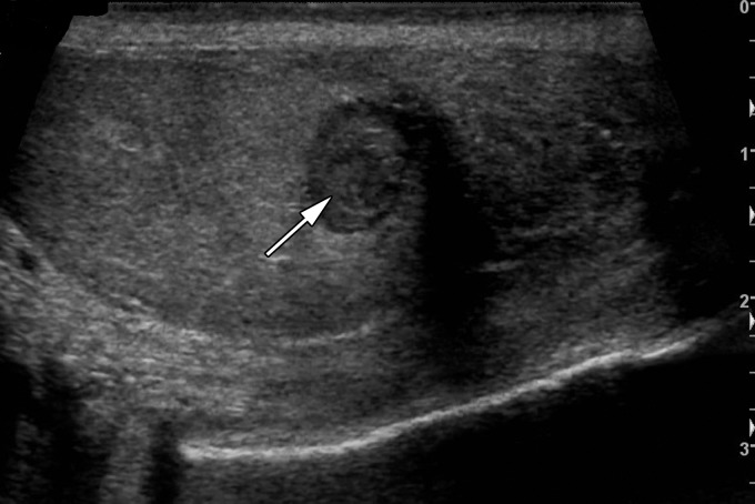
Figure 8a.
Testicular hematoma in a 29-year-old man with scrotal pain after trauma. (a, b) Gray-scale US (a) and color Doppler (b) images of the left testis show a well-circumscribed hypoechoic lesion (arrow in a) without color Doppler flow (arrow in b), compatible with testicular hematoma. (c) Color Doppler image 10 days later shows the lesion to be smaller and more hypoechoic (arrow), characteristic of testicular hematoma.

