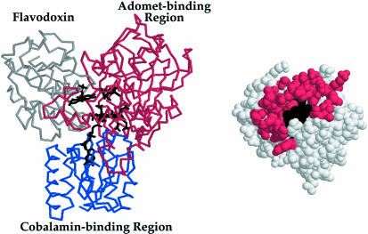
Figure 5.
Modeled interface of flavodoxin with methionine synthase. The FMN, cobalamin, and AdoMet cofactors of flavodoxin (11) (gray), the cobalamin-binding region (31) (blue), and AdoMet-binding domain (27) (red) of methionine synthase are juxtaposed for electron transfer from FMN to cobalamin and for methyl transfer from AdoMet to cobalamin according to a published docking model (28). Predicted contacts of flavodoxin with the AdoMet-binding domain are mapped onto the surface of flavodoxin, which is shown in the same orientation as in Fig. 3.

