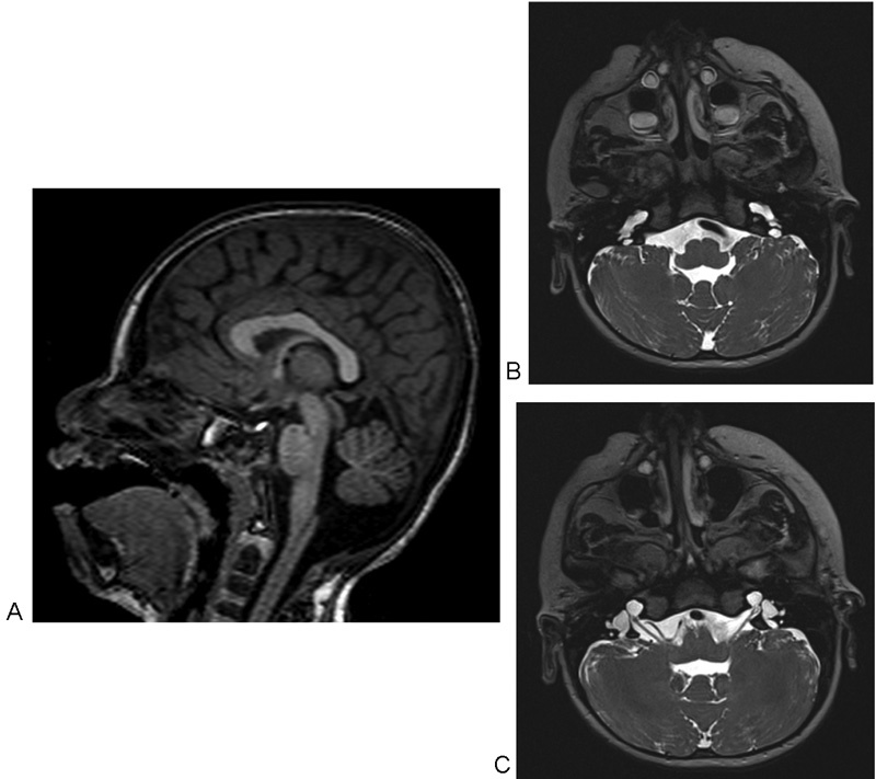
Fig. 1.

Brain magnetic resonance imaging at 4 months of life. ( A ) Cerebellar vermis hypoplasia. ( B ) Bilateral dilatation of the endolymphatic sac, and dilatation of the cochlear basal turn. ( C ) Internal acoustic canals enlargement and agenesis of their osseous fundus, agenesis of the modiolus, dilation of vestibules and vestibular aqueducts, and cystic enlargement of the medial and apical turns, which appear fused.
