
Figure 4. PHD proteins are functionally redundant in T lymphocytes.
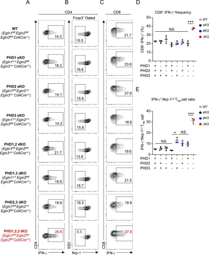
(A–B) Phenotype of pulmonary CD4+ T cells in mice with indicated PHD protein deficiency. Representative flow cytometry of IFN-γ expression (A) and Nrp-1 expression by Foxp3+ Treg cells (B).
(C–D) IFN-γ expression by pulmonary CD8+ T cells in mice with indicated PHD protein deficiency. Representative flow cytometry (C) and replicate values (E) are shown.
(E) Ratio of CD4+ IFN-γ+ Teff cells to Nrp-1Lo Foxp3+ Treg cells of total pulmonary CD4+ T cells in mice with the indicated PHD protein deficiency.
Data are representative of ≥ 2 independent experiments with ≥ 3 mice per genotype. Bars and error represent mean ±SEM of replicate measurements. *P<0.05, ***P<0.001 (Student’s t-test).
See also Figure S4.