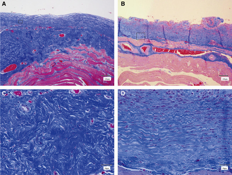
Fig. 2.

Masson trichrome staining. A, Low-power field image of a capsule formed around a smooth expander prosthesis (scale bar = 100 μm). The surface of the capsule is smooth. B, Low-power field image of a capsule formed around a textured expander prosthesis (scale bar = 100 μm). The surface of the capsule is textured. C, Enlarged field image of the square in A (scale bar = 30 μm). D, Enlarged field image of the square in B (scale bar = 30 μm).
