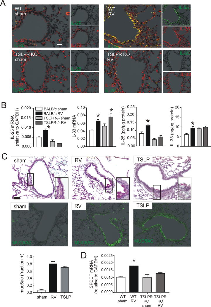
FIG 6.
Innate cytokine expression in immature TSLPR KO mice after RV infection. Six-day-old BALB/c or TSLPR KO mice were inoculated with sham or RV. A. Two days after infection, lungs were stained for IL-33 (red) and IL-25 (green; bar is 50 µm). B. Lung mRNA and protein expression were measured 2 days after infection (N=3–5, mean±SEM, *different from sham, one-way ANOVA). C. Six-day-old BALB/c mice were inoculated with sham, RV, or recombinant TSLP (rTSLP). Lung sections were prepared 3 weeks after infection and stained with PAS solution or anti-Muc5ac. Fractional airway epithelial staining for Muc5ac was measured (N=3, mean±SEM, * different from sham, p<0.05, one-way ANOVA). D. Lung SPDEF mRNA was measured 2 days after RV infection. (N=4, mean±SEM, *different from sham, one-way ANOVA).

