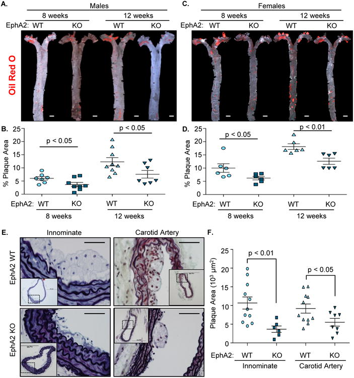
Figure 1. EphA2 deletion reduces plaque size in multiple vascular beds.

A-D) Plaque formation in mice fed Western diet for 8 or 12 weeks was determined following Oil Red O staining. Representative micrographs are shown (A/C), and plaque area was calculated as a percent of the aorta staining positive for Oil Red O (B/D). n = 7-9 male, 6 female. Scale bar = 1 mm E, F) Male Apoe-/- controls (EphA2 WT) and EphA2-/-Apoe-/- (EphA2 KO) mice were fed Western diet for 8 weeks, and the innominate and carotid arteries were stained with Movat pentachrome stain. Plaque area was quantified as the neointimal area within the internal elastic lamina. n = 6-11 innominate artery, 8-11 carotid sinus. Scale bar = 100 μm. Data are expressed as mean ± SEM. Student's T-tests were used for statistical comparisons.
