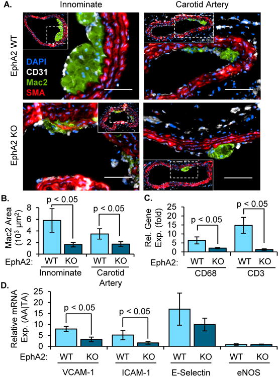
Figure 2. Reduced inflammation in early atherosclerotic plaque formation.

Male EphA2 WT) and EphA2 KO mice were fed Western diet for 8 weeks. A,B) Macrophage area (Mac2 positive, green) was determined by immunohistochemistry in both the innominate (n = 6-10) and carotid arteries (n = 14-21). Smooth muscle (SMA-positive, red) and endothelial (CD31-positive, white) cell staining are also shown. Quantification of average macrophage area (B) is provided. Scale bar = 100 μm. C/D) Relative gene expression between atherosclerosis-prone (aortic arch (AA)) and protected (thoracic aorta (TA)) regions was determined by mRNA isolation and qRT-PCR and compared between EphA2 WT and EphA2 KO mice. Expression of (C) macrophage (CD68) and T cell (CD3) marker genes or (D) markers of endothelial activation (VCAM-1, ICAM-1, E-selectin) were normalized to the housekeeping genes PPIA and Rpl13a. n = 5-8. Data are expressed as mean ± SEM. Student's T-tests were used for statistical comparisons.
