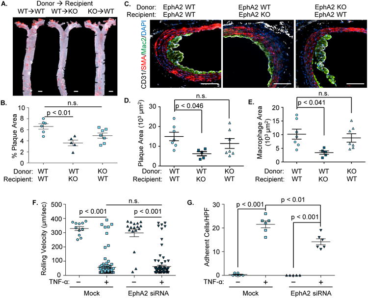
Figure 4. EphA2 deletion from resident cells but not hematopoietic cells confers atheroprotection.

A-E). EphA2 WT and EphA2 KO mice were irradiated and received either EphA2 WT or KO bone marrow cells. Four weeks post-irradiation, mice were fed Western diet for 12 weeks. Atherosclerotic plaque formation was assessed in (A/B) the aorta by Oil Red O staining and (C/D/E) the innominate artery by immunohistochemistry for macrophages (Mac2, green), smooth muscle (SMA, red), and endothelial cells (CD31, white). Scale bar = 1 mm (A) or 100 μm (C). n = 5-7. D) Plaque area was quantified as the neointimal area within the internal elastic lamina. E) Macrophage area was quantified as the total area of positive Mac2 staining. F/G) Human aortic endothelial cells with or without EphA2 knockdown (siRNA) were treated with TNF-α (10 ng/mL, 4 hours) and labeled primary human monocytes were perfused over the endothelial monolayer under physiological flow. Rolling velocity and firmly adherent monocytes were analyzed per high powered field averaged over six fields. n = 3 in duplicate. Data are expressed as mean ± SEM, Statistical comparisons were performed using One-way (B-E) or Two-way ANOVA (F, G) with Bonferroni post-tests.
